page 1
(The Study of Threes)
http://threesology.org
Visitors as of 11/18/2019
| Pages in the Biological & Physiological 3s Series: | ||
| B/P 3s pg. 1 | B/P 3s pg. 2 | B/P 3s pg. 3 |
| B/P 3s pg. 4 | B/P 3s pg. 5 | B/P 3s pg. 6 |
| B/P 3s pg. 7 | B/P 3s pg. 8 | B/P 3s pg. 9 |
| B/P 3s pg. 10 | B/P 3s pg. 11 | The Devil's Advocate and 3s Research |
Let us of the present day (circa early 21st century) acknowledge that we are in a very early stage of humanity's cognitive evolution, and that human physiology retains much of its primivity, despite the mind's desire to move beyond its primitive beginnings and attachments. Like an infant whose umbilical cord is severed so that it may grow and learn to run free; humanity seeks to severe its mind from the spinal cord so that it too can grow and run free. As such, we use different models, different techniques by which to count with to provide an account of who we are (or at least think we are), where we are, where we came from, and what's "out there", which the limitations of our bodies constrain us from exploring and that no present day form of artificiality (drugs, alcohol, religion, music, etc...) has enabled an honest, not habituating, non-maligning means of exploration. Hence, when we give an "account" of a subject, we are providing a counted itemization of ideas, of impressions, of imagination... whether or not a particular person uses an explicit effort to display their account with an easily detectable enumeration. Nonetheless, information is serialized, just as one counts numbers or expresses the alphabet.
Note: Ensembles of "Threes" in Anatomy can be found here:

- Dr. McNulty's List of Three's in Anatomy
- Validity of the Rule of Threes and Anatomical Relationships in the Thoracic Spine by Clayton K Oakley, Sarah A Keim Janssen, Joseph P Pankratz, Travis L McCumber, Kevin D Treffer, Anthony B Olinger
When we speak of biology some readers may necessarily include the study of genetics, philosophy, comparative anatomy, pharmacology, psychology (modern and ancient forms), witchcraft/sorcery/alchemy (antiquities of thought in History), medicine, bio-chemistry, chemistry and atomic structure as well; since they incorporate an "origins" interest as part of there overall research model which includes various "asides" like desserts accompanying a main meal... because they incorporate an idea of a Universal inter-connectedness. In the 1960s the everyday common approach was to use a dichotomous theme of macro/micro towards unraveling the mysteries of different subjects. Very often one might find an extension of dichotomous orientation by inserting the Eastern views of Yin and Yang (with the later adoption of including the notion of complementarity, unity, oneness, etc.... While some came to view the yin and yang model as being too simplistic, though medical partitioners in China became adept at using such a "crude" system of cataloguing and vantage point, others choose to adopt a mathematics approach— thinking it to be superior... that is, dealing with a more finely tuned structure of observing and working with fundamentals; most people apparently often overlook the basic structure of Mathematics as advancing a different means of cataloguing dichotomies as well.
However, before touching more on this, let me first provide those who are interested in using an "origins" or basic fundamental structures approach, with a short list of trichotomies involving Nature applicable to biology. The initial illustration came from Mark Mahin's: Nature Loves the Number Three. The present illustration depicts a few other examples I added on:

Clearly we can't have biology without chemistry nor chemistry without atomic particles and such particles without some sort of spatial configuration (such as a Universe) to put them in.
Yet let me step back to provide some rather instructive information about mathematics since I believe it to be a Westernized model of the Easternized Yin/Yang perspective; both of which are illustrating the use of a binary form of conceptualization from different vantage points and elaborations of symbolic characterizations. And let me further note that from the earlier binary proportions associated with the Yin/Yang idea linked to the binary model of perceived sexuality (Yin is female and Yang is male); there was a later trend to conceptualize beyond the binary towards a trinary with the adoption of the I-Ching with its proposed Triads, which... when looked closely at, are actually embellished biads, because the "stick" configurations used as a form of tally sheet, does not illustrate a stick figure with three lines. Whereas we have a so-called triad with 1 and 2 lines that are repeated, we do not have an actual figure where there is a 1-, 2-, 3- line configuration.
Similarly, when we look at Mathematics and make note of the very many dichotomies being used as a basic structure, and effort to move beyound the "two"... this binary into a three... a trinary, is likewise deprecated to small efforts seen in such ideas as Trigonometry (sine-cosine-tangent), set theory (1,2,3...), Pythagorean theorem (A2 + B2 = C2, and Boolean logic (And, Or, Not)). In other words, both the Eastern born Yin/Yang ideology and the Western born Mathematics perspective, are fundamental expressions of human conceptualization using a binary formula as a philosophy for cataloguing and organizing observations. They are different models of counting which have elaborated the primitive's usage of learning to count in a (three-patterned) "1- 2- many" formula. It is the same pattern we can recognize being used by Nature. Atomic physics is replete with "threes" just as biology is.
Both the Eastern-born Yin/Yang and Western born Mathematics illustrate a similar type of brain functioning taking place in different eras. Note also that the Yin and Yang model of dichotomization does not have multiple branches of consideration while the Mathematics model of dichotomization does. This may well be due to the multiplicity of language variations encountered in the West for migration, wars, commerce, religion, etc... The different branches of Mathematics is like encountering a community of different speakers selling different goods, though they themselves may think they engage in a common lingua franca. Nonetheless, both are elaborations of a basic type of thinking that is striving to explore beyond its fundamental patterning. A good example is to look at the present day usage of a binary code for computers which some are striving to supplant (let us say augment) by adopting a three-patterned trinary (ternary) system. It is a counting system much like that encountered long ago by primitive hominids who were learning to count. The mental functioning of the human brain appears to repeat its earlier steps, albeit in embellished ways. The lack of development in this respect may be due to the presence of a "conservation of number" as a result of humans being subjected to an incrementally deteriorating environment which requires the use of small numbers as a survival mechanism. Hence, while the triplet code in DNA may (or may not) have an internalized capacity of growing (numerically) beyound its repeating "three" pattern, it is being forced to comply with the requirements of surviving in a deteriorating environment. Indeed, if DNA is part of Evolution, why isn't it evolving in real/actual terms and not merely imaginative terms of supposition someone might want to concoct to be argumentative?


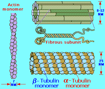
The cytoskeleton is a network of 3 types of protein polymers, namely the microtubules, the intermediate filaments and the microfilaments. It also contains numerous proteins that are associated with the fibrous polymers. They nucleate, bundle, cap and link the fibers with each other, with cell organelles and the plasma membrane. It is the mechanical and functional framework for every known cellular function.
Microtubules ~ Intermediate Filament ~ Actin Filaments
 ---Northwestern.edu Glossary --- |
 |
Fibrous proteins that occur in the cytoplasm, referred to as the cytoskeleton maintain the shape of the cell as well as anchoring organelles, moving the cell and controlling internal movement of structures. Microtubules function in cell division and serve as a "temporary scaffolding" for other organelles. Actin filaments are thin threads that function in cell division and cell motility. Intermediate filaments are between the size of the microtubules and the actin filaments. | |
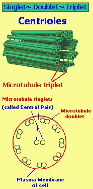
In doublet and triplet microtubules the wall of one microtubule partly consists of the wall of an attached microtubule. (In other words, there is a type of overlapping... or, if you will... a type of fusion. Just like the Sun's three "moments" [dawn- noon- dusk] that will one day "fuse" into 1 as the Sun enlarges and the Earth's rate of rotation slows.) |
 |
The image at right represents singlet, doublet, and triplet microtubules in cross section. The A tubule of
a doublet or triplet microtubule is a complete cylinder, made of 13
protofilaments. "Piggyback" B or C tubules are made of less than 13
protofilaments, usually 10. |
 |
3 primary cellular energy molecules:
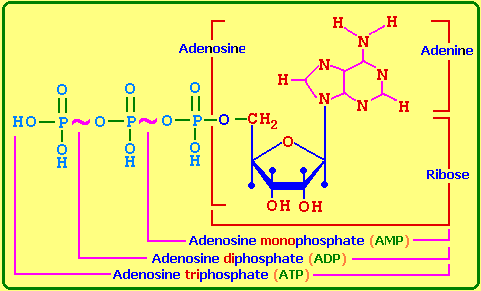
- AMP = Adenosine- Mono- Phosphate: (Mono = one)
- ADP = Adenosine- Di-
Phosphate: (Di = two)
- ATP = Adenosine- Tri- Phosphate (Tri = three)
- 3 general categories of Eukaryotes: Animals ~ Fungi ~ Plants
- 3 units of measurement for microscopic organisms: Micrometres ~ Nanometres ~ Ångstroms
- 3 groups of basic cells: Eubacteria ~ Archaebacteria ~ Eukaryotes
- 3 germinal layers: Endoderm ~ Mesoderm ~ Ectoderm
- 3 sequenced organism development: "Monoploblastic" ~ Diploblastic ~ Triploblastic
- 3 types of molecular bond: Covalent ~ Ionic ~ Polar covalent
- 3 isomerism types: Structural (Ethyl alcohol ~ Geometric (Maleic acid) ~ Optical (L-Lactic acid)
- 3 hydrocarbon chain types: Straight (Propane) ~ Branched (Isobutane) ~ Circular (Cyclopropane)
- 3 main fatty acid categories: Saturated ~ Monounsaturated ~ Polyunsaturated
*** Omega-3 family of Essential Fatty Acids is said to be needed for optimum health
Omega-3 fatty acids come in three main types:
- Eicosapentaenoic (EPA)
- Docosahexaenoic (DHA)
- Alpha-linolenic acid (ALA), which doesn't have the proven benefit for the heart as EPA and DHA.
- 3 basic chemical reaction substances: Acids ~ Bases ~ Salts
- 3 layers of Arteries, Capillaries, Veins: Connective tissue ~ Muscle ~ Endothelium
Specifically, the common structural pattern that can be seen in all blood vessels with the exception of capillaries is:
|
The Tunica Intima Delimits the vessel wall towards the lumen of the vessel and comprises its endothelial lining (typically simple, squamous) and associated connective tissue. Beneath the connective tissue, we find the internal elastic lamina, which delimits the tunica intima from:... The Tunica Media Is formed by a layer of circumferential smooth muscle and variable amounts of connective tissue. A second layer of elastic fibers, the external lastic lamina, is located beneath the smooth muscle. It delimits the tuenica media from:... The Tunica Adventitia Consist mainly of connective tissue fibres. The tunica adventitia blends with the connective tissue surrounding the vessel. The definition of the outer limit of the tunica adventitia is therefore somewhat arbitrary. |

|
Note: In Capillaries, only the tunica intima is present, which typically consists of:
- The Endothelium-
- Its basal lamina-
- And an incomplete layer of cells surrounding the capillary, the pericytes. Pericytes have contractile properties and can regulate blood flow in capillaries.
Three types of capillaries can be distinguished based on features of the Endothelium:
|
Continuous capillaries are formed by "continuous" endothelial cells and basal lamina. The endothelial cell and the basal lamina do not form openings, which would allow substances to pass the capillary wall without passing through both the endothelial cell and the basal lamina. Both endothelial cells and the basal lamina can act as selective filters in continuous capillaries. Fenestrated capillaries The endothelial cell body forms small openings called fenestrations, which allow components of the blood and interstitial fluid to bypass the endothelial cells on their way to or from the tissue surrounding the capillary. The fenestrations may represent or arise from pinocytotic vesicles which open onto both the luminal and basal surfaces of the cell. The extent of the fenestration may depend on the physiological state of the surrounding tissue, i.e. fenestration may increase or decrease as a function of the need to absorb or secrete. The endothelial cells are surrounded by a continuous basal lamina, which can act as a selective filter. Discontinuous capillaries Are formed by fenestrated endothelial cells, which may not even form a complete layer of cells. The basal lamina is also incomplete. Discontinuous capillaries form large irregularly shaped vessels, sinusoids or sinusoid capillaries. They are found where a very free exchange of substances or even cells between bloodstream and organ is advantageous (e.g. in the liver, spleen, and red bone marrow). |

|

| |
 |
Three types of lymph vessels can be distinguished based on their size and morphology:
Lymph capillaries
Are somewhat larger than blood capillaries and very irregularly shaped. They begin as blind-ending tubes in connective tissue. The basal lamina is almost completely absent and the endothelial cells do not form tight junctions, which facilitates the entry of liquids into the lymph capillary. Temporary openings in the endothelial lining of the lymph capillaries also allow the entry of larger particles into the lymph capillaries (lipid droplets, which are absorbed from the lumen of the gut do not enter blood capillaries, but enter the circulation via lymph vessels which are found in the villi of the ileum and jejunum). Lymph capillaries merge to form:...
Lymph collecting vessels
Which are larger and form but otherwise appear similar to lymph capillaries. The lymph is moved by the compression of the lymph vessels by surrounding tissues. The direction of lymph flow is determined by the valves. Lymph vessels empty intermittently into lymph nodes from which the lymph continues in efferent lymph vessels.
Only very little lymph is returned from the limbs if they are immobilized, which illustrates the importance of muscular action in lymph transport. This is also the reason for immobilizing limbs that are either infected or that have been bitten by venomous Australians. The effect can also be observed after long inter-continental flight when you may feel that your shoes and socks are just about one number too small. Finally, impeded lymph drainage is one of the problems associated with surgery which requires the removal of lymph nodes and which thereby interrupts the lymph collecting vessels.
Eventually the lymph collecting vessels merge to form:...
Lymph ducts
Which contain one or two layers of smooth muscle cells in their wall (some textbooks call this layer the tunica media of lymph vessels). They also form valves. The walls of the lymph ducts are less flexible in the region of the attachment of the valves to the wall of the duct, which may give a beaded appearance to the lymph ducts. Peristaltic contractions of the smooth muscle contribute to the movement of lymph towards the heart in addition to the compression of the ducts by surrounding tissues.
The largest lymph duct of the body, the thoracic duct, drains lymph from the lower half and upper left quadrant of the body and empties the lymph into the circulation by merging with the vascular system close to the junction of the left internal jugular and subclavian veins. That it is the largest lymph duct does not mean that it is a large vessel when compared to the large arteries and veins. It actually is not much larger (about 5mm in diameter) than one of the superficial forearm veins.
Three different (general) "types" of arterial vessels may be distinguished, but it must not be forgotten that these so-called types occur as part of a continuous, gradually changing, vascular morphology based on functional requirements:
- Large elastic (conducting) arteries, which leave the heart and are continuous with...
- Medium and small muscular (distributing) arteries, which join...
- Arterioles, which are continuous with capillary vessels.
Arterioles have a relatively thick muscular wall in comparison to their luminal diameter; the lumen of the smallest arterioles can accommodate about three to four red blood corpuscles.
From the structural/functional point of view, elastic tissue is the most important component in the larger vessels, whereas smooth muscle is the most important in the smaller vessels.
Three "types" of veins according to size:
- Venules
- Small and medium-sized veins
- Large veins
Venules can be recognized when they are about 20 µm in diameter (about three red corpuscles across) and possess an endothelial lining, a thin layer of collagenous fibers with some fibroblasts. They have neither muscle fibers nor elastic fibers.
At 10 weeks of development there are two veins and one artery in the umbilical cord. Pg. 136, LIFE by Lennart Nilsson, Hardcover ISBN # 0-8109-5842-2
3 last steps of blood coagulation:
- Inactive factor X... Active factor X...
- Prothrombin... Thrombin...
- Fibrinogen... Fibrin
3 closures occurring as the neonate begins to breathe on its own:
- Closure of ductus arteriosus
- Closure of umbilical artery and vein
- Closure of ductus venous
The following is a three-to-one variation:
|
If the skin of the forearm of many individuals is lightly stroked with a blunt instrument, a white line appears at the site of the stroke within 20 seconds. The blanching becomes maximal in about 30 to 40 seconds and then gradually disappears within 3 to 5 minutes. |
|
A triple response occurs if the skin is stroked more strongly with a sharp pointed instrument:
|
Other variations of the 3 to 1 ratio can be seen starting on this page:
- 3 substances metabolized for energy needs: Carbohydrates ~ Fats ~ Proteins
- Triglycerides are the main storage forms of fatty acids.
- 3 dietary monosaccharides well absorbed: Glucose ~ Galactose ~ Fructose.
- 3-patterned general formula for carbohydrates: Cx(H20)y
|
Respired Carbohydrate Catabolism = 3 stages Glucose is the most common carbohydrate energy source used by cells. The respiration of glucose typically occurs in three principal stages:
Fermented Carbohydrate Catabolism = 1 stage The initial stage of fermentation usually is also Glycolysis. However, once glycolysis has taken place, pyruvic acid is converted into one or more different products, depending on the type of cell. Unlike respiration, there is no Krebs Cycle or Electron Transport Chain in fermentation. Accordingly, the ATP yield is much lower. |
3 basic metabolic types:
- The P-type in which the (PNS) Parasympathetic Nervous System dominates
- The S-type with a dominating (SNS) Autonomic Nervous System
- A balanced type.
3 factors combine to regulate our metabolism: Nutrition ~ Glandular system ~ Nervous system.
3 main food components: Glucose (carbohydrates) ~ Amino acids (proteins) ~ Fatty acids (fats).
3 directions are possible after glycolysis.It is here that most intracellular energy problems develop. Pyruvic acid can either combine with carbon dioxide and become oxaloacetic acid, or it may lose carbon dioxide and form acetyl coenzyme A. (Glucose is normally the most important fuel molecule. Through several reactions along the glycolytic pathway it is split in half to form pyruvic acid. Niacinamide and vitamin B1, as co-enzymes, are the necessary vitamins at this stage; magnesium is required in addition. This glycolysis occurs anaerobically.)
Carbohydrate Metabolism:
- 1st stage: Glycolysis, the oxidation of glucose to pyruvic acid. The enzymes of glycolysis catalyze the splitting of glucose, a six-carbon sugar, into two 3-carbon sugars.
- 2nd stage: {3 common labels}; Krebs Cycle, Citric Acid Cycle, Tricarboxylic Acid Cycle. All three carbon atoms in pyruvic acid are released as CO2.
- 3rd stage: Electron Transport Chain.
Three classes of carrier molecules in Electron transport chains:
- Flavoproteins
- Cytochromes
- Ubiquinones
The chemiosmotic mechanism of ATP (Adenosine- TRI- Phosphate) synthesis, called Chemiosmosis, was first proposed by the British Biochemist Peter Mitchell in 1961. Although it took several years to gain full acceptance, it is now recognized as a major milestone in the history of biochemistry. The three steps are:
|
| Source/Method = (1~ 2~ 3) |
ATP Yield | Misc. Value {666) |
#1. Oxidation of glucose to pyruvic acid = Method: Substrate-level phosphorylation #2. Production of 2NADH = Method: Oxidation phosphorylation in electron transport chain |
2ATP 6ATP |
6 = 3 x 2 |
|
#1. Formation of acetyl CoA produces 2 NADH = Method): Oxidative phosphorylation in electron transport chain |
6ATP |
6 = 6 x 1 |
#1. Oxidation of succinct CoA to sluicing acid = Method: Equivalent of ATP; substrate-level phosphorylation #2. Production of 6NADH = Method): Oxidative phosphorylation in electron transport chain) #3. Production of 2 FADH = Method: Oxidative phosphorylation in electron transport chain) |
2GTP 18ATP 4ATP |
18 = 6 x 3 |
Note: In the foregoing table I indicate the presence of a 1~ 2~ 3 occurrence as well as providing a bit of numerical correlation for those who are numerologically-inclined. My intent is merely to present the same values on one side of the "equation" which are denoted by the # sign and the other side of the table (equation) which again reflect the 1~ 2~ 3. Heaven forbid that the end result is 666! Our carbohydrate metabolism is in league with the Devil! (But anyone who has been on a diet knows this... the devil made you eat that last cookie.)
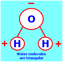
3
molecules to water: 2 hydrogen and 1 oxygen B.C. comic strip by Johnny Hart, ©Field Enterprises Inc. 1972 |
 |
3 divisions to the world's water ("blood") supply:
- Ocean = 97%
- Ice sheets, glaciers, ice fields, ice floes = 2%
- Steams, lakes, rivers, ground water, soils = 1%
(Different sources give different percentages but there are generally 3 categories.)
3 flow pattern types of Pulmonary Circulation:
- Zone A (Alveolar pressure exceeds intra vascular pressure).
- Zone B (Alveolar pressure intermediate between arteriolar and velar pressures).
- Zone C (Intra vascular pressure exceeds alveolar pressure)
3 principal forms of carbon dioxide (CO2) carried in the blood:
- In physical solution
- As bicarbonate ions
- As crazing compounds.
3 minerals (in homeostasis) that are essential for health and life: Calcium ~ Phosphate ~ Magnesium
3 classes of Amines (ammonia compounds):
- Primary, NH2R
- Secondary, NHR2
- Tertiary, NR3
Various Biological/Physiological 3s:
- 3 classes of Neurons: Motor (Monopolar) ~ Sensory (Bipolar) ~ Inter (Multipolar)
- 3 main cellular components of Blood: Red blood cells ~ White blood cells ~ Platelets
- 3 blood plasma protein groups: Globulin ~ Albumin ~ Fibrinogen
- 3 white blood cell (leucocyte) groups: Granulocyte ~ Lymphocyte ~ Monocyte
- 3rd corpuscle: alternate name for platelets
- Band 3 protein is complex molecule in red blood cell (erythrocyte) membranes
- 3 letters (and combinations thereof) used for blood typing: (A~ B~ 0)
- 3 main blood channels: Veins ~ Arteries ~ Capillaries
- 3 interferon types: (White blood cells) ~ (Fibroblasts-connective tissue) ~ (Lymphocytes)
- 3 carbon-to-carbon bonds: Single (Ethene)~ Double(Ethylene)~ Triple (Acetylene)
- 3 genotypes: AA (homozygous dominant) ~ aa (homozygous recessive) ~ Aa (heterozygous)
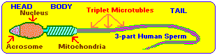
3 part general division to the human sperm: (Head ~ Body ~ Tail)
*** Since the fertile life of the human egg cell lasts at most one day and that of the human sperm at most two days, there is a period of about three days during which copulation can result in conception (the day when the egg is fertile and the two preceding days).
- Generally speaking, the egg traverses the fallopian tube in about 3 days
- Implantation of the egg in the uterus occurs in about 3 days.
- Sperm can travel at a rate of 3 inches per hour.
- A spermagonium gives rise to 3 active cells and 1 resting cell.
As an aside note: Whenever I see a picture of a sperm I get the impression of a kite with a tail, flying overhead some distance away. However, when I was younger, I once pictured a kite I was flying, as a spider and the cross-stick braces as part of the hour-glass figure seen on black-widow spiders. As such, this image began to unravel into a sort of story in which I saw myself as a fly and the string was part of the spider's web. This impression was so life-like and amusing that it influenced me to write a poem shortly thereafter, that I had not thought about for some years.
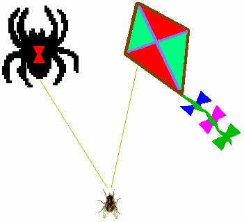 A kite is a spider...
releasing a silken web and I am a fly- er, unaware of the potential dread. While religious minds may see a cross I can clearly see an hourglass or a figure "8," at a different toss, the mind's labyrinth passage... some fear to pass To and fro at a busy pace encircling the sky with clouds as if it were in a sort of race, and we on Earth the cheering crowds. All in all to claim the prize the Sun, a chalice, of golden ore encasing its glow from peering eyes, as the sky grows dark the more. But I do not heed the tugging line steadfast I hold, captured thus wind or not, tis mine! All mine! amidst other fly- ers, and their fuss. There I stand, or so I think thinking tis I who know the better quite unaware the sky did shrink, from a single cloud, the web, my fetter. Too tightly gripped, I won't let go the loss of such is very frightening... and others say, they told me so, that a spider can strike like lightning. |
3 types of eggs classified according to their yolk concentration:
| Egg Type | Description | Examples |
| Isolecithal, alecithal |
Having a small amount of yolk distributed in the cytoplasm; or lacking visible yolk. | Sea urchins, Humans |
| Teloecithal | Having the yolk more highly concentrated at the vegetal pole than at the animal pole; cytoplasm concentrated toward the animal pole. | Moderately telolecithal: Frogs,
Salamanders Strongly telolecithal: Birds, Reptiles, some Sharks |
| Centrolecithal | Having the yolk located in the center of the egg and surrounded by cytoplasm. | Insects |
3 cell surface changes during the formation and maturation of the egg:
Dramatic changes in the cell surface occur during the growth phase of oogenesis. Most striking is the formation of an external coat of polysaccharide-rich material known as the vitelline envelope in non-mammalian oocytes or the zona pellucida in mammalian oocytes. This external coat, which protects the egg from chemical and physical injury, may remain intact well past the time of fertilization. when the external envelope is produced by the oocyte itself, it is referred to as a primary coat. When formed by the follicle cells it is called a secondary coat, and when produced by the oviduct or other maternal tissue, it is termed a tertiary coat.
The appearance of these various layers differs significantly among species. Especially variable is the tertiary layer, which ranges from the jelly coat of amphibian eggs to the egg white and shell of hens' eggs. Impervious shells of this latter type must of course be added to the egg after fertilization, lest they prevent entry of sperm.
In non-mammalian oocytes the biochemical and morphological changes that occur during the growth phase of oogenesis impart an obvious asymmetry (polarity) to the developing oocyte.
In other words, more primitive life forms exhibit a characteristic pattern-of-two... giving rise to the idea that there may not only be a recurring 1 - 2- 3 maturational development taking place, but that which promoted such many billions of years ago during evolutionary stages may still be identifiable and replicable.
3 standard text reproductions of the miniature homunculus:
- Hartsoeker's little man with a big head neatly curled up within a sperm.
- Dalen Patius' tiny animals.
- D'Agoty's homunculus suspended in a glass of water and visible "even without the help of lenses."
Sandor Fuenczi had claimed that the erotic genital secretion of females has an oceanic basis: The odor of the vagina comes from the same substance (Tri-methlamine) as the decomposition of fish gives rise to. (And to think that for centuries some men have been claiming that there is something fishy about women which makes them difficult to understand.)

- 3 trimester divisions in pregnancy
- 3 bones of the pelvic girdle: Ilium ~ Pubis ~ Ischium
- 3 stages of labor: Dilation of the cervix ~ Delivery of the baby ~ Delivery of the placenta
- 3 stages to ovarian follicle development
- 3 layers to female uterus: Perimetrium ~ Myometrium ~ Endometrium
- 3 embryonic somites: Dermatomes ~ Myotomes ~ Sclerotomes
- 3 membranes surround embryo: Amnion ~ Chorion ~ Allantois
- 3 days after birth is a baby's customary first check-up
- 3 months old is when most babies begin shedding tears
Prolactin is a protein hormone principally concerned with stimulating breast development and milk production in women. It is considered that it may play some role in reproduction. It is a single-chain protein with 198 amino acids and three disulfide bridges.
3 distinct phases to a triphasic curve:
- The first phase of lower temperatures before ovulation (follicular phase).
- The second phase of higher temperatures after ovulation (corpus luteum phase). At ovulation the basal body temperature rises and stays up because of the progesterone hormone which is being produced by the corpus luteum and which increases the bbt.
- The third phase where the bbt curve again rises to a third level (triphasic) of temperatures about a week or so after
ovulation.
The triphasic curve supposedly shows implantation, but there is no sufficient scientific evidence to prove or disprove it.
A "perfect" Triphasic case illustrated in the following chart. The triphasic shift is clearly visible and is confirmed by a light spotting.
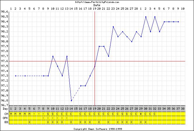
While this case is said to be very rare, and that the triphasic pattern is generally masked if it is even present, the fact remains that the idea of a "perfect three-patterned" state is representative of the presence of some underlying cognitive three-patterned orientation, formulation, process.
Initial posting: Wednesday, 18th November 2019... 7:26 AM
Updated Posting: Tuesday, October 25th, 2021... 6:00 AM
Latest Update: Friday, February 24th, 2023... 11:16 AM
Herb O. Buckland
herbobuckland@hotmail.com